Copyright © 2025 Open Medicine Foundation. All Rights Reserved.
To explore the hypothesis that low-grade neuroinflammation in the central nervous system can be part of the cardinal symptoms of ME/CFS.
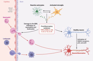
The overall aim is to explore the hypothesis that low-grade neuroinflammation in the central nervous system can be part of the cardinal symptoms of ME/CFS. Accumulating evidence suggests that ME/CFS patients can suffer from chronic neuroinflammation, the sustained activation of microglia and astrocytes, to strongly influence neurocognitive disturbances, headache, and fatigue and contribute to its progression. A major question is whether inhibition of the inflammatory response has the ability to reverse or slow down its symptoms. Furthermore, for many other brain diseases, such as neurodegenerative disorders, neuroinflammation is emerging as a cause, rather than a consequence, of the pathogenesis.
Neuroinflammation can possibly be monitored in cerebrospinal fluid (CSF) as a result of the inflammatory reaction itself or as secondary markers of cell degradation and impaired cell repair mechanisms. Our study will be the first to systematically measure CNS structural abnormalities in a large ME/CFS case-control study using CSF collection and in-situ measurement of intracranial pressure together with the study of microglia activation by the application of a novel diffusion-based MRI methodology. Added to this will be collected biospecimens to perform extensive state-of-the-art analyses with the ambition to identify diagnostic and prognostic biomarkers.

OMF is a non-profit 501(c)(3) organization
(EIN# 26-4712664). All donations are tax-deductible to the extent allowed by law.


Open Medicine Foundation®
29302 Laro Drive, Agoura Hills, CA 91301 USA
Phone: 650-242-8669
info@omf.ngo
Copyright © 2025 Open Medicine Foundation. All Rights Reserved.
What are the advantages of giving from your Donor Advised Fund (DAF)?
How do I make a donation through my DAF?
Just click on the DAF widget below. It is simple and convenient to find your fund among the over 900 funds in our system.
Still can’t find your fund?
Gifting of Stock
Broker: Schwab
DTC #: 0164
Account #: 47083887
Account Registered as:
Open Medicine Foundation
29302 Laro Drive
Agoura Hills, CA 91301
Please speak to your personal tax advisor and then email or call OMF at 650-242-8669 to notify us of your donation or with any questions.